1.INTRODUCTION
This is the reactions that occur in the body tissue as the result of an injury or irritant. The irritant could be traumatic, bacterial, degenerative or even regenerative. If the severity of the injury causes destruction of tissue (necrosis) then the inflammatory reaction will occur in the surrounding area. The inflammatory process is the defensive and is an attempt to remove the irritant, debris and dead cells. During the initial phase, inflammation is very essential as it acts as the chief defence mechanism and promotes healing.
The most common inflammations which occurs around the elbow :
a) Tennis elbow (Lateral epicondylitis)
b) Golfer’s elbow (Medial epicondylitis)
c) Student (Bursitis) elbow
a. Tennis elbow:
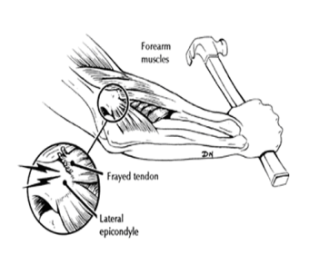
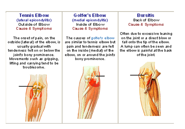
Tennis Elbow or Lateral Epicondylitis is a condition when the outer part of the elbow becomes painful and tender, usually as a result of a specific strain, overuse, or a direct bang. Sometimes no specific cause is found.
b.Golfer’s elbow:
Golfer’s elbow or medial epicondylitis is an injury to the muscles and tendons on the medial aspect of the elbow. It is more common in throwers and golfers hence the ‘nicknames’.
These problems, tennis elbow and golfer’s elbow, are both forms of tendonitis. Tendons are the ends of muscles that attach to bone. Because of the force of the muscle, the points of insertion of the tendon on the bone are often
pointed prominences. The medical names of Tennis Elbow (lateral epicondylitis) and Golfer’s Elbow (medial epicondylitis) come from the names of these bony prominences where the tendons insert, and where the inflammation causes the pain.
c. Student (bursitis) elbow:
Below the tip of the elbow is a sack of fluid called a bursa. A bursa is used to help lubricate the movement of tissues in joints. However if you take a hard impact onto the back of the elbow or are constantly leaning on the elbows the bursa can become inflammed or bleeding can occur in the bursa. The result is a painful swelling on the bony bit at the back of the elbow.
2.ANATOMY
The joints and muscles of the elbow complex are designed to serve the hand. They provide mobility for the hand in space by apparent shortening and lengthening of the upper extremity. Rotation at the elbow complex provides additional mobility for the hand. In conjunction with providing mobility for the hand, the elbow complex structures also provide stability for skilled or forceful movements of the hand when performing activities using tools or implements.
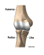
The elbow complex consists of the elbow joint (humeroulnar and humeroradial articulations) and the proximal and distal radioulnar joints. The apparent shortening and lengthening of the upper extremity occur at the elbow joint, which is formed by the distal end of the humerus and the proximal ends of the radius and ulna.
The elbow complex is made of three bones, three ligaments, two joints and one capsule. The joining (articulation) of the humerus with the ulna and radius is commonly called the elbow joint. The elbow joint is a uniaxial hinge joint that allows only flexion and extension. There is about 1450 of flexion measured from the 0-degree position of extension. The 00 of extension is when arm is completely straight.
2.1 Articular Surfaces
The elbow-joint is a ginglymus or hinge-joint. The trochlea of the humerus is received into the semilunar notch of the ulna, and the capitulum of the humerus articulates with the fovea on the head of the radius. The articular surfaces are connected together by a capsule, which is thickened medially and laterally, and, to a less extent, in front and behind.
The joint (articulation) between the radius and the ulna is known as the radioulnar joint. These two bones connect with each other at both ends. At this end the top (head) of the radius pivots within a groove in the ulna (radial notch). This joint is a pivot joint, which is uniaxial, and allows only the turning of your forearm clockwise (supination) and counterclockwise (pronation). There should be about 90 degrees of supination and 80 degrees of pronation. When pronation or supination occur, the radius moves around the ulna. The ulna does not move.
Jointcapsule:
The humeroulnar and humeroradial joints and the superior radioulnar joint are enclosed in a single joint capsule. The joint capsule attached anteriorly to the proximal attachment of the capsule is just above the coronoid and radial fossae, and distally it is inserted into the ulna on the margin of the coronoid process and into the annular ligament. Laterally, the capsule’s attachment to the radius blends with the fibers of the lateral collateral ligament. Medially, the capsule blends with fibers of the medial collateral ligament. Posteriorly the capsule is attached to the humerus along the upper edge of the olecranon fossa. The capsule is large and loose.
Capsule of elbow-joint (distended). Anterior aspect:
Capsule of elbow-joint (distended). Posterior aspect.
Anterior and Posterior ligaments.
Medial and lateral collateral ligaments
Anterior Ligament:
The anterior ligament is a broad and thin fibrous layer covering the anterior surface of the joint. It is attached to the front of the medial epicondyle and to the front of the humerus immediately above the coronoid and radial fossæ below, to the anterior surface of the coronoid process of the ulna and to the annular ligament being continuous on either side with the collateral ligaments. Its superficial fibers pass obliquely from the medial epicondyle of the humerus to the annular ligament. The middle fibers, vertical in direction, pass from the upper part of the coronoid depression and become partly blended with the preceding, but are inserted mainly into the anterior surface of the coronoid process. The deep or transverse set intersects these at right angles. This ligament is in relation, in front, with the Brachialis, except at its most lateral part.
The Medial or Ulnar Collateral Ligament (ligamentum collaterale ulnare; internal lateral ligament)
Is triangular shaped and spans the inside (medial) of the elbow. Medial means towards your body, so the medial side of your elbow, would be the side facing your body. This ligament is a thick triangular band consisting of two portions, an anterior and posterior united by a thinner intermediate portion.
The anterior portion, directed obliquely forward, is attached, above, by its apex, to the front part of the medial epicondyle of the humerus; and, below, by its broad base to the medial margin of the coronoid process.
The posterior portion, also of triangular form, is attached, above, by its apex, to the lower and back part of the medial epicondyle; below, to the medial margin of the olecranon. Between these two bands a few intermediate fibers descend from the medial epicondyle to blend with a transverse band which bridges across the notch between the olecranon and the coronoid process. This ligament is in relation with the Triceps brachii and Flexor carpi ulnaris and the ulnar nerve, and gives origin to part of the Flexor digitorum sublimis.
The Anterior band anterior portion of Medial Collateral Ligament provide 300, 600 and 900of elbow flexion. Posterior band of anterior portion Medial Collateral Ligament provide 1200 pf elbow flexion. Posterior portion of medial colleteral ligament provide 55,85 and 900 of flexion to full flexion.
The Lateral (Radial) Collateral Ligament:
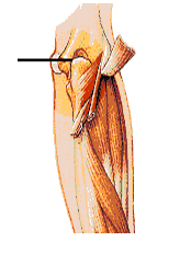
Is also triangular shaped and attaches on the lateral side of your humerus (lateral epicondyle) and on the annular ligament and the lateral side of the ulna. Lateral means away from your body, so the lateral side of your elbow would be the side facing away from your body. These two ligaments provide a great deal of medial and lateral stability to the elbow. It is weaker but more elastic than Medial Collateral Ligament. This ligament is a short and narrow fibrous band attached, above, to a depression below the lateral epicondyle of the humerus; below, to the annular ligament, some of its most posterior fibers passing over that ligament, to be inserted into the lateral margin of the ulna. It is intimately blended with the tendon of origin of the Supinator.
The Posterior Ligament:
This posterior ligament is thin and membranous, and consists of transverse and oblique fibers. Above, it is attached to the humerus immediately behind the capitulum and close to the medial margin of the trochlea, to the margins of the olecranon fossa, and to the back of the lateral epicondyle some
little distance from the trochlea. Below, it is fixed to the upper and lateral margins of the olecranon, to the posterior part of the annular ligament, and to the ulna behind the radial notch. The transverse fibers form a strong band which bridges across the olecranon fossa; under cover of this band a pouch of synovial membrane and a pad of fat project into the upper part of the fossa when the joint is extended. In the fat are a few scattered fibrous bundles, which pass from the deep surface of the transverse band to the upper part of the fossa. This ligament is in relation, behind, with the tendon of the Triceps brachii and the Anconæus.
Left elbow-joint, showing anterior and ulnar collateral ligaments
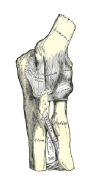
posterior and radial collateral ligaments

Relations of elbow joint:
Anterior: Brachialis, Median nerve, Brachial artery, Tendon of Biceps
Posterior: Triceps, Anconeus
Medial: Ulnar nerve, Flexor carpi ulnaris, Common flexors
Lateral: Supinator, Extensor carpi radialis brevis, Other common extensors
2.4 MUSCLES:
The muscles in relation with the joint are, in front, the Brachialis; behind, the Triceps brachii and Anconeus; laterally, the Supinator, and the common tendon of origin of the Extensor muscles; medially, the common tendon of origin of the Flexor muscles.
FLEXORS
Nine muscles cross the anterior aspect of elbow joint but only three of these muscles (Brachialis, Brachioradialis, Biceps) have primary functions. Remaining six muscles (pronator Teres, flexor carpi radialis, flexor carpi ulnaris, flexor digitorum superficialis, and palmaris longus)
Brachialis:
It arises from the anterior surface of the lower portion of the humeral shaft and attaches by thick broad tendon to the ulnar tuberosity and coronoid process. This muscle is supplied by musculo cutaneous nerve .This muscle is very strong elbow flexor .
Biceps:
The Biceps muscle has two heads, a long and short head. Both heads arises from the scapula.
v The long head arises from the supraglenoid tubercle and runs over the head of the humerus and out of the joint capsule to descent through the bicipital groove to joint with the short head.
v The short head arises as a thick flat tendon from the coracoid process of scapula.
The muscle fibers arising from the two tendons unite in the middle of the upper arm to form the prominent muscle bulk of the upper arm. Muscle fibers from both heads insert by way of strong flattened tendon on rough posterior area of tuberosity of radius. Other fibers of biceps brachi insert into bicipital aponeurosis that extends medially to blend with the facia that lies over forearm flexors. This is supplied by musculocutaneous nerve (C5,C6) it acts as a strong supinator when forearm flexed. Short head for flexion of arm and long head to prevent upward displacement of head of humerus.
Brachioradialis:
Is originates from the lower third of lateral supracondylar ridge of humerus and inserts onto the distal end of radius just proximal to radial styloid process. Because of it’s more lateral attachment, it is more effective as an elbow flexor when the forearm is in neutral position.
Common extensors of elbow:
Triceps:
It has 3 heads
- Long head
- Medial head
- Lateral head
Long head crosses both the glenohumeral joint at the shoulder as well as elbow joint. The long head arises from the infraglenoid tubercle of the scapula by a flattend tendon that blends with the glenohumeral joint capsule.
Medial head cross only the elbow joint. Medial head covers an extensive area as it arises from entire posterior surface of humerus.
Lateral head arises from narrow ridge on the posterior humeral surface.
These 3 heads inserts into the olecronon process of humerus. Supplied by radial nerve (C7,8) .It is powerful extensor of elbow.
Anconeus:
It originated from the lateral epicondyle of humerus and inserts into both olecronon process and adjacent posterior surface of ulna.
2.5 Arteries:
The arteries supplying the joint are derived from the anastomosis between the profunda and the superior and inferior ulnar collateral branches of the brachial, with the anterior, posterior, and interosseous recurrent branches of the ulnar, and the recurrent branch of the radial. These vessels form a complete anastomotic network around the joint.
2.6 Nerves:
Joints receive branches from
- Ulnar nerve
- Median nerve
- Radial nerve
- Musculo cutaneous nerve through it’s branch to the brachialis.
2.7 Movements:
Common movements of elbow joint
- Flexion
- Extension
Flexion is produced by the action of the Biceps brachii and Brachialis, assisted by the Brachioradialis and the muscles arising from the medial condyle of the humerus; extension, by the Triceps brachii and Anconæus, assisted by the Extensors of the wrist, the Extensor digitorum communis, and the Extensor digiti quinti proprius.
Carrying Angle:
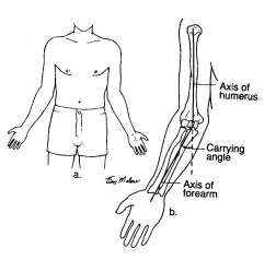
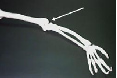
The transvers axis of the elbow joint is directed medially and downwards. Because of this, the extended forearm is not straight line with the arm, but makes an angle of about 1630 with it. This is known as carrying angle. The factors responsible for production of the carrying angle are
- Medial flange of the trochlea is 6mm deeper than the lateral flange.
- The superior articular surface of the coronoid process of ulna is placed oblique to the long axis of bone.
2.8 Injuries
The elbow joint will be used in most all upper body exercises and movements. A common injury is tendonitis felt on the back of the elbow. Most people assume that this is their triceps tendon. However, in most cases, it is the tendon from the wrist flexor muscles. These sets of muscles originate from the rear side of the elbow (medial epicondyle of the humerous) and insert onto the fingers in different arrangements. During lying triceps extensions (skull crushers), some will allow there wrist to be bent backwards. This will cause stress and chronic pain to the back side of the elbow. While most think it is their elbow hurting, it is in reality the tendons from their hands, due to poor grip. This can be fixed by maintaining a strong grip on the bar and keeping your wrist flexed or stiffened. If you have this injury, and feel pain here during lying triceps curl, stay away from exercises for the triceps, when arm is over your head. If any other exercise causes pain, stay away from it for at least four to six weeks. Use cables or dumbbell kickbacks, while working your triceps. After this period of time your tendon should be healed enough to start back using light weight. Remember to keep a firm and correct grip on the bar.
Tennis elbow is another common malady affecting many people. This is an overuse syndrome or tendonitis involving the outside region of the elbow (lateral humeral epicondyle). In simple terms it hurts on the back and/or outer side of the elbow. Microscopic tears of these tendons are very common for people in their forties, and it may or may not have come from athletics. You can test yourself by noting any tenderness over the elbow. Also you should be able to elicit tension and pain when the elbow is extended, your forearm is turned counter clockwise (pronate) and the wrist is flexed. If you feel pain when doing this, then you should layoff of any activity that causes pain. Such as throwing motions, some triceps exercises, carrying heavy objects with that arm and anything else that may affect the area.
Tendons in general take a long time to heal, because of the lack of blood flow to the area. Tendons naturally have a low blood supply, so the nutrients it needs for healing doesn’t get there as fast as it would for a muscle. So using light resistance with high repetitions will force blood into the working muscle, and allow more blood flow to get to the injured tendon.
3.Bio-Mechanics
Study of Mechanics in the human body is refered to as bio-mechanics. Bio-Mechanics consists of area of
- Kinetics
- kinematics
Kinematics is the area of bio-mechanics which include description of motion without regard for the forces producing the motion.
Kinematic variables for movement include
- Type of motion that occurring
- Location of movement
- Direction of motion
- Magnitude of motion
- Rate and duration
Kinetics is the area of bio-mechanics concerned with the forces producing motion or maintaining equilibrium.
3.1 Axis of Motion:
The axis for flexion and extensdion is relatively fixed and passes through the center of the trochlea and capitulum bisecting the longitudinal axis of the shaft of the humerus. When the upper extremity is in the anatomic position, the long axis of the humerus and the long axis of the forearm ofrm an acute angle medially when they meet at the elbow. The angulation is due to the configuration of the articulating surface and results in a normal valgus angulating of the forearm in relation to the humerus . This angle is called the carrying angle and is slightly greater in women than men. The average angle in men is about 50 whereas in women it is about 100to 150. An increase in the carrying angle is considered to be abnormal, especially if it occurs unilaterally, when the angle is increased beyond the average, it is called cubitus valgus.
Normally, the carrying angle disappear when the forearm is pronated and the elbow is in full extension and when the forearm is flexed against the humerus in full elbow flexion. Carrying angle did not change during the range of elbow flexion/extension.
3.2 Range of Motion:
A number of factors determine the amount of motion that is available at the elbow joint. These factors include the type motion position of forearm and the position of the shoulder. The range of active flexion at the elbow is usually less than the range of passive motion, because the bulk of contraction flexors on the anterior surface of the humerus interferes with the approximation of the forearm with the humerus. The active range of motion for elbow flexion considered to be from about 135 degrees to 145 degrees, whereas the range for passive flexion is between 150 and 160 degrees. The position of the forearm also effect the Range Of Motion. When the forearm is in either pronation or midway, between supination and pronation, the Range Of Motion is less than it is when the forearm is supinated. The position of the shoulder may affect the Range Of Motion available to the elbow. The bony components MCL and anterior joint capsule contribute equally to resist valgus stress in full extension. The bony components provide one half of the resistance to varus stress in full extension and the lateral collatral and joint capsule provide other half of resistance.
Approximation of coronoid process with the coronoid fossa and of the rim of the radial head in radial fossa limits extreme of the flexion. In 90 degrees of flexion the anterior part of MCL provide the primary resistance to both distraction and valgus stress. If the anterior portion of the MCL becomes lax through over stretching, medial instability will result when the elbow is in flexed positions. Also the carrying angle will increase. Majority of the resistance to varus stress when the elbow is flexed to 90 degrees is provided by the osseous structures of the joints and only a slight amount by the LCL and the joint capsule. The anterior joint capsule provide only slightly to varus/valgus stability and provides little resistance to destraction when the elbow is flexed. Co-contraction of the flexors and extensors muscles of the elbow , wrist, hand help to provide stability for the elbow during forceful motions of the wrist, fingers and in activities in which the arms are used to support the body weight.
4. Pathology
Inflammation is best defined as the local reaction of vascularized tissue to injury. CORNELIUS CELSUS described the four cardinal signs of inflammation:
v Rubor (redness)
v Tumor (swelling),
v Calor (heat)
v Dolor (pain)
JULIUS COHNHEIM (1839-1884) who provided one of the first microscopic descriptions of the inflammatory process.
4.1 Types of Inflammation:
The inflammatory response has two themes :
v Inflammation
v Repair
I. Inflammation:
is of two types:
v Acute
v Chronic
Acute Inflammatory Process:
In acute inflammation the intensitive of the reaction is determined by both the severity of the injurious agent and the reactive capability of the host. The intensity and duration of the inflammatory reaction depends on the precarious balance between the strength of the attacker vs., that of the host. Depending on the severity of the injury and the adequacy of the defence, the inflammation may remain localized to it’s site of origin or may evoke systemic responses.
The local clinical signs of inflammation are the heat, redness, swelling and pain immortalized by CELSUS. A fifth clinical sign, loss of function was added by VIRCHOW. The local heat and redness results from dilatation of the micro circulation in the environs of the injury. The swelling is largely produced by the escape of fluid, plasma proteins and cells from the blood into the perivascular tissues. Here pain can be induced by the prostaglandins when there is a simultaneous release of bradykinin or serotonin or when there is increased tissue tension due to edema.
The escape of fluid, proteins, and cells from the vascular system is know as exudation. An exudates is inflammatory extra vascular fluid that has a high protein concentration, much cellular debris, and specific gravity above 1.020. A transudate is a low-protein fluid with a specific gravity of less than 1.012.
The local manifestations of acute inflammations clearly highlight the three major components of the inflammatory response.
- Changes in vascular flow and caliber
- Changes in vascular permeability
- Leukocytic exudation
4.2 Haemodynamic Changes:
These changes are best observed in thin, transparent injured tissues under microscope. Observable changes are
- First there is transient vasoconstriction of arterioles
- Vasodilatation, which first involves the arterioles and then result in opening of new capillaries and venular beds in the area. Thus comes about the increased blood flow. This is the hallmark of early Haemodynamic changes in the acute inflammation and the cause of the heat and redness.
- Slowing of the circulation follows, brought about by the increased permeability of the microvasculature, with the outpouring of the protein-rich inflammatory fluid into the extravascular tissues. This results in stagnation or “statis”.
Sir THOMOS LEWIS discovered “triple response”. Lewis pointed out that when the skin of the forearm of a normal individual is firmly stroked by a blunt instrument, such as the tip of the lead pencil or the edge of the ruler, three separate changes can be observed.
v First, within seconds, a dull redline develops along the line of the stroke
v Second a bright red hallow appears about the stroke mark.
v Third feature appears-swelling (wheal) accompanied by balancing of the original stroke mark.
4.3 Changes in the vascular permeability:
Leakage of fluid as a consequence of changes in the permeability of the micro vasculature, with the resultant tissue swelling (edema), is a major and constant characteristic of all acute inflammatory reactions. Increased vascular permeability and exudation of plasma proteins, the mark of acute inflammatory edema.
 B. Acute inflammation B. Acute inflammation |
-
- Normal hydrostatic pressure of about 32mm hg at arterial end of capillary and 12mm of hg at venous end . Mean capillary pressure equals colloid oncotic pressure.(horizontal line)
- Acute inflammation . Mean capillary pressure is increased because of arteriolar dilatation, while oncotic pressure is reduced because of increased permeability of vascular wall. Result is net excess of extravasated fluid.
According to STARLING’S hypothesis, the normal fluid balance is maintained by two opposing sets of forces. Those that cause the fluid to move out of circulation are the osmotic pressure of the interstitial fluid and the intravascular hydrostatic pressure; those that cause fluid to move in are the osmotic pressure of plasma proteins and the tissue hydrostatic pressure. The balance of these forces is such that there is net small movement of fluid outwards, but this flud normally drains into the lymphatics, and no edema occurs. Factors that tend to either increase intravascular hydrostatic pressure or decrease intravascular osmotic pressure will result in increased movement of fluid out of the capillary and the formation of edema. In inflammatory edema there is a loss of high-protein fluid due to a leaky endothelium and therefore a reduction of the intravascular osmotic pressure, accompanied by increased osmotic pressure of the interstitial fluid leading to impairment of the return of the fluid to the blood on the venous end of the capillary. There is thus marked out flow of fluid.
Chronic Inflammation:
Persistent inflammatory stimuli leads to chronic inflammation. It occurs in three ways:
- It may follow acute inflammation because of the persistence of the inciting stimulus
- It may be due simply to repeated bouts of acute inflammation, with the patient showing successive attacks of fever, pain, swelling.
- More curiously, it may begin insidiously as a low grade, smoldering response that never acquires the classic features of acute inflammation.
II. Repair:
Repair is may be by the formation of :
v Homogenous tissue: Where the new tissue are exactly like the original tissue, e.g., bone, fibrous tissue and epidermal structures.
v Scar tissue: the original tissue is replaced by fibrous tissue during the process of repair.
5.General Clinical Features of Inflammation
The common clinical features of inflammation:
5.1 Acute Inflammation:
- Pain is present over a diffuse area due to pressure on the nerve endings.
- The pain is present even during rest and is aggravated by activity; it may be reffered to other areas from the site of primary inflammation.
- Skin and the tissue temperature is raised at the site of the lesion with marked tenderness, effusion, swelling and redness. This occurs due to increased blood flow due dilatation of capillaries.
- There is increased capillary permeability with the leakage of protein and plasma. The tissue fluid with high degree of fibrinogen seeps out into the adjacent tissue, giving rise to swelling.
- Passive range of motion may be painful and restricted due to pain and protective spasm in the antagonistic group of muscles.
- There may be a temporary loss of function of the affected area, depending upon the degree and site of inflammation.
5.2 Chronic Inflammation:
- Pain is usually absent during the rest. The pain may be present over a localized area near the site of lesion and is aggravated by specific activities which results in stretching of the inflamed area.
- There is presence of granulation tissue.
- The skin temperature is not raised; effusion is absent; tenderness, if at all present, is minimal.
- Passive range of motion is restricted but is not painful.
- The pain is elicited only when the joint is stretched to the point of its restriction. This restriction of ROM is usually due to tendon shortening, adhesions or capsular fibrosis in the soft tissues.
6.COMMON INFLAMMATIONS
TENNIS ELBOW
Definition:
Tennis elbow is a painful elbow disorder. This term is misleading because most people who have it did not get it from playing tennis. In fact, tennis elbow seldom has any connection with fun and games. Also called as Epicondylalgia or Epicondylitis. It is an extra articular condition believed to be caused by strain or incomplete rupture of the forearm extensor muscles.
The technical name for tennis elbow is “lateral epicondylitis”. This term indicates an inflammation occurring near a small point or projection of the upper arm bone (humerus) just above the elbow joint on the outer side of the arm. However, pain can also occur in other areas of the forearm and elbow. Some experts suggest that “lateral elbow pain syndrome” is a more accurate name, but this term is not yet commonly used.
The pain from tennis elbow comes mainly from injured or damaged tendons near the elbow. Tendons are strong bands of tissue that connect muscles to bones. When repeatedly stressed or overused, tendons can become inflamed. This results in a painful condition called tendonitis, the medical term for inflammation of a tendon. Tennis elbow is simply a specific type of tendonitis that occurs in a particular part of the elbow.
Tennis elbow is an injury to the muscles and tendons on the outside (lateral aspect) of the elbow that results from overuse or repetitive stress. The narrowing of the muscle bellies of the forearm as they merge into the tendons create highly focused stress where they insert into the bone of the elbow.
Muscles arising from lateral aspect of elbow
Extensors of the forearm:
Superficial Muscles arising from lateral epicondyle
Anconeus:
It is originating from lateral epicondyle of the humerus and inserted into lateral aspect of olecranon process of ulna and Upper one fourth of posterior surface of ulna supplied by radial nerve (C7,8;T1). It acts as a weak extensor of the elbow.
Brachioradialis:
It is originating from upper 2/3rd of lateral supracondylar ridge of humerus and lateral intermuscular septum and get inseted into lateral side of radius just above the styloid process supplied by radial nerve (C5,6,7). It acts as flexor of forearm.
Extensor carpi radialis londus:
It is originating from lower 1/3rd of lateral supracondylar ridge of humerus and from the lateral intermuscular septum and get inserted into base of second metacarpal bone supplied by radial nerve (C6,7). It acts as extension and abduction of wrist.
Extensor carpi radialis brevis:
Extensor digitorum:
It takes origin from lateral epicondyle and the muscle ends in a tendon which splits into 4 parts, one for each digit other than thumb. Over the proximal phalanx the tendon for each digit divides into 3 slips- one intermediate and 2 collateral. The intermediate slip is inserted into the dorsal aspect of the base of the middle phalanx . The collateral slips reunite to be inserted into dorsal aspect of base of distalphalanx . Supplied by posterior interosseous nerve (C7,8). It’s action is extension of Inter phalangeal and Metacarpo phalangeal and wrist joint.
Extensor digiti minimi:
It takes origin from common extensor origin and get inserted into dorsal aspect of the base of the middle phalanx and base of distal phalanx and supplied by posterior interosseus nerve(C7,8). It acts as extensor of little finger at interphalangeal and metacarpophalangeal joints.
Extensor carpi ulnaris:
It takes origin from common extensor origin and posterior border of ulna and get inserted into medial side of the base of the fifth metacarpal bone supplied by posterior interosseus nerve (C7,8). It’s action is extension of wrist and adduction of the hand.
Deep muscles:
v Supinator
v Abductor pollicis longus
v Extensor pollicis brevis
v Extensor pollicis longus
v Extensor indicis
Incidence:
More than 50% of recreational tennis players suffer from lateral epicondylitis. Peak incidence is in the 4th and 5th decades of life.
History:
It was described from Writer’s cramps by RANGE in 1873. It was MADRIS who called it as Tennis Elbow.
Athletes complain of insidious-onset lateral elbow pain exacerbated by inciting activities. Acute-on-chronic pain is suggestive of a frank rupture of the extensor origin.
Mechanism of Injury:
Injury to the lateral aspect of the elbow is the most common upper extremity tennis injury. Tennis elbow is generally caused by overuse of the extensor tendons of the forearm, particularly the extensor carpi radialis brevis. Commonly experienced by the amateur player, this injury is often a result of (1) a one-handed backhand with poor technique (the ball is hit with the front of the shoulder up and power generated from the forearm muscles), (2) a late forehand swing preparation with resulting wrist snap to bring the racquet head perpendicular to the ball, or (3) while serving, the ball is hit with full power and speed with wrist pronation (palm turned downward) and wrist snap which increases the stress on the already taught extensor tendons.
The development of tennis elbow can often be traced to the way of using the forearm muscles. These muscles control hand and wrist movements. They are attached to tendons that connect them to only two small points of bone just above the elbow, one on the outer side, the other on the inner side.
Muscles connected to the outer side of the elbow are responsible for:
- straightening the fingers,
- bending the wrist upwards,
- rolling the forearm into a palms-up position.
There are weak points in the way tendons connect these muscles to the bone above the elbow. The points where the tendons attach are sometimes too small to handle the strong force of the powerful muscles. These tendons can get overloaded when the hand and forearm are used in strong, jerky movements such as gripping, lifting, or throwing.
Tendons do not stretch when pulled. They are rope-like structures made of strong, smooth, shiny fibers. Strong forces or sudden impacts, however, can eventually tear their fibers apart in much the same way a rope becomes frayed. This type of injury is called a strain, and usually results in formation of scar tissue. Over time, strained tendons become thickened, bumpy, and irregular. Without rest and time for the tissue to heal, strained tendons can become permanently weakened.
Most experts believe it is due to the small tears that develop in the tendons. Other possible causes include the development of scar-like tissue under the tendon, wear and tear of the elbow joint, or irritation and inflammation of nerves that pass near the elbow region.
Etiology:
Commonly implicated sports include tennis, racquet ball, squash and fencing.
Although called tennis elbow, lateral epicondylitis is much more commonly seen in people who are over using their arm doing something else. It could equally well be called “plasterer’s elbow” or “mechanic’s elbow” or “painter’s elbow”.
Rarely the inflammation comes on without any definite cause, and this may be due to an arthritis, rheumatism or gout. Sometimes the problem is partly or completely due to a neck problem, which is causing pain in the elbow via the nerves from the neck.
Risk factors :
The development of tennis elbow often relates to the way that workers carry out activities such as gripping, twisting, reaching, and moving. These activities can become hazardous when they are done:
- In fixed or awkward position,
- With constant repetition,
- With excessive force, and
- Without allowing the body to recover from the wear and tear.
Tennis elbow is associated with jobs that require repeated or forceful movements of the fingers, wrist, and forearm. It can develop because of too much force at once or small amounts of force for too long a period.
Specific movements associated with the development of tennis elbow include:
- Simultaneous rotation of the forearm and bending of the wrist,
- Stressful gripping of an object in combination with inward or outward movement of the forearm,
- Jerky, throwing motions,
- Movements to hit objects with the hand.
The movements in the first two activities mentioned above (rotation, bending, and gripping) are particularly hazardous when done while the arms are extended forward and/or sideways away from the body (torso).
Pathology of Tennis elbow:
The damage that tennis elbow incurs consists of tiny tears in a part of the tendon and in muscle coverings. After the initial injury heals, these areas often tear again, which leads to hemorrhaging and the formation of rough, granulated tissue and calcium deposits within the surrounding tissues. Collagen, a protein, leaks out from around the injured areas, causing inflammation. The resulting pressure can cut off the blood flow and pinch the radial nerve, one of the major nerves controlling muscles in the arm and hand.
Tendons, which attach muscles to bones, do not receive the same amount of oxygen and blood that muscles do, so they heal more slowly. In fact, some cases of tennis elbow can last for years, though the inflammation usually subsides in 6 to 12 weeks.
Signs and Symptoms :
Tennis elbow can cause extreme tenderness on the outer side of the elbow. This tenderness becomes painful when the wrist and elbow are moved in certain ways. These include:
- Bending the wrist while straightening the elbow
- Trying to straighten the wrist against resistance while straightening the elbow,
- Trying to bend the hand back against resistance while straightening the elbow, and
- Trying to straighten the fingers against resistance.
- Swelling over the origin of the extensor tendon.
- Pain is persistent and gives raise to continuous annoyance, it radiates down the forearm. There is a sense of weakness when attempts are made to perform lifting movements.
In a medical examination, pain experienced in any three of these movements can indicate the possibility of tennis elbow. Usually there is no outward sign of redness or swelling. Most often tennis elbow affects only one arm, usually the one that does most at work.
Tennis elbow can appear in many different ways. Some people get symptoms gradually after doing the same type of work for several years. Others get it suddenly, soon after they start doing a new type of work. Sometimes it can develop immediately following a single violent muscle exertion or after an elbow becomes injured. In other cases, tennis elbow occurs for no obvious reason.
Prevention:
- Discontinue or modify the action that is causing the strain on your elbow joint. If you must continue, be sure to warm up for 10 minutes or more before any activity involving your arm, and apply ice to it afterward. Take more frequent breaks.
- Try strapping a band around your forearm just below your elbow. If the support seems to help you lift objects such as heavy books, then continue with it. Be aware that such bands can cut off circulation and impede healing, so they are best used once tennis elbow has disappeared.
GOLFER’S ELBOW
Definition:
This is a similar injury to tennis elbow only it affects the inside of the elbow instead. It is more common in throwers and golfers hence the ‘nicknames’. Also known as flexor / pronator tendinopathy this injury is seen in tennis players who use a lot of top spin on their forehand shots.
It should be kept in mind that elbow epicondylitis is not limited to those persons playing tennis, golf, baseball or swimming and can result from any activity that puts the lateral or medial compartments of the elbow under similar repetitive stress and strain (e.g., hammering, turning a key, screw driver use, computer work, excessive hand shaking).
Common muscles arising from the medial epicondyle:
Flexors of Forearm:
Pronator teres:
Muscle is attached on the medial side of the humerus (medial epicondyle). This muscle also originates from the ulna (coronoid process). It crosses the elbow joint on the front surface (anterior), and runs diagonally to insert on the lateral surface of the radius at about midpoint. Its function is to turn your forearm counter clockwise (pronation), and is assistive in elbow flexion.
Flexor carpi radialis :
Is taking origin from the medial epicondyle of the humerus and get inserted into palmar surface of the bases of the second and third metacarpal bones which is supplied by median nerve. The main action is flexion of the wrist and abduction of the wrist.
Palmaris longus:
Is taking origin from medial epicondyle of humerus get inserted into distal half of flexor retinaculum and the apex of the palmar aponeurosis which is supplied by median nerve. The main action is flexion of the wrist.
Flexor carpi ulnaris:
Taking origin from the medial epicondyle of humerus and from the medial margin of the olecranon, and from the posterior border of the ulna get inserted into pisiform bone which is supplied by ulnar nerve. Main action is flexion of wrist, adduction of the wrist.
Flexor digitorum superficialis:
Taking origin from medial epicondyle of humerus and from the anterior border of the radius and some fibres arise from fibrous arch passing from the ulna to radius and connecting the two heads. The muscle ends in four tendons one each for medial four fingers which is supplied by medial nerve main action is flexion of the proximal interphalangeal joints.
Incidence:
Medial epicondylitis is approximately 1/5th as frequent as it’s lateral counter part.
History:
Athletes presents with insidious, activity-related medial elbow pain and swelling. Question the player about the participation in at risk sports. Provocative activities include the acceleration phase of throwing, the tennis serve, and forehand racquet strokes.
Mechanism of Injury:
Medial epicondylitis is less common and characteristically occurs with wrist flexor activity and pronation. Medial epicondylitis can result from (1) late forehand biomechanics where the player quickly snaps the wrist to bring the racquet head forward, (2) the back-scratch or cocking phase when serving, which places tremendous stress on the medial tissues of the elbow, (3) in the right elbow of a right-handed golf swing by throwing the club head down at the ball with the right arm rather than pulling the club through with the left arm and trunk (also referred to as “golfers elbow”), or (4) improper pulling technique with certain swim strokes, especially the backstroke (also referred to as “swimmers elbow”).
Etiology:
By far the most common cause of golfers elbow is overuse. Any action which places a repetitive and prolonged strain on the forearm muscles, coupled with inadequate rest, will tend to strain and overwork those muscles.
There are also many other causes, like a direct injury, such as a bump or fall onto the elbow. Poor technique will contribute to the condition, such as using ill-fitted equipment, like golf clubs, tennis racquets, work tools, etc. While poor levels of general fitness and conditioning will also contribute.
Risk Factors:
The development of golfers elbow often relates to the way that workers carry out activities such as gripping, twisting, reaching, and moving. These activities can become hazardous when they are done:
- in fixed or awkward position,
- with constant repetition,
- with excessive force, and
- without allowing the body to recover from the wear and tear.
Pain is produced by extension of elbow, supination and valgus strain. It can develop because of too much force at once or small amounts of force for too long a period.
Pathology:
The damage that golfers elbow incurs consists of tiny tears in a part of the tendon and in muscle coverings. After the initial injury heals, these areas often tear again, which leads to hemorrhaging and the formation of rough, granulated tissue and calcium deposits within the surrounding tissues. Collagen, a protein, leaks out from around the injured areas, causing inflammation. The resulting pressure can cut off the blood flow and pinch the median nerve, one of the major nerves controlling muscles in the arm and hand.
Tendons, which attach muscles to bones, do not receive the same amount of oxygen and blood that muscles do, so they heal more slowly. In fact, some cases of golfers elbow can last for years, though the inflammation usually subsides in 6 to 12 weeks.
Signs and Symptoms:
v Pain is the most common and obvious symptom associated with golfers elbow. Pain is most often experienced on the inside of the upper forearm, but can also be experienced anywhere from the elbow joint to the wrist.
v Weakness, stiffness and a general restriction of movement are also quite common in sufferers of golfers elbow. Even tingling and numbness can be experienced.
v Pain on the inside of the elbow when you grip something hard.
v Pain when wrist flexion (bending the wrist palm downwards) is resisted.
v Pain on resisted wrist pronation – rotating inwards (thumb downwards).
v Pain caused by lifting or bending the arm or grasping even light objects such as a coffee cup.
v Difficulty extending the forearm fully (because of inflamed muscles, tendons and ligaments).
v Tenderness and pain at the medial epicondyle.
Prevention:
There are a number of preventative techniques which will help to prevent golfers elbow, including bracing and strapping, modifying equipment, taking extended rests and even learning new routines for repetitive activities
Firstly, a thorough and correct warm up will help to prepare the muscles and tendons for any activity to come. Without a proper warm up the muscles and tendons will be tight and stiff. There will be limited blood flow to the forearm area, which will result in a lack of oxygen and nutrients for the muscles. This is a sure-fire recipe for a muscle or tendon injury.
Before any activity be sure to thoroughly warm up all the muscles and tendons which will be used during your sport or activity.
Secondly, flexible muscles and tendons are extremely important in the prevention of most strain injuries. When muscles and tendons are flexible and supple, they are able to move and perform without being over stretched. If however, your muscles and tendons are tight and stiff, it is quite easy for those muscles and tendons to be pushed beyond their natural range of movement. When this happens, strains, sprains, and pulled muscles occur. To keep your muscles and tendons flexible and supple, it is important to undertake a structured stretching routine.
And thirdly, strengthening and conditioning the muscles of the forearm and wrist will also help to prevent golfers elbow. There are a number of specific strengthening exercises you can do for these muscles.
OLECRANON BURSITIS (MINOR’S ELBOW)
Definition:
Bursitis simply put is the inflammation of a bursa. In the normal state, the bursa provides a slippery surface that has almost no friction. problem arises when a bursa becomes inflamed. The bursa loses its gliding capabilities, and becomes more and more irritated when it is moved. When the condition called bursitis occurs, the slippery bursa sac becomes swollen and inflamed. The added bulk of the swollen bursa causes more friction within already confined spaces. Also, the smooth gliding bursa becomes gritty and rough. Movement of an inflamed bursa are painful and irritating.
Repeated trauma to the posterior aspect of the elbow joint is the precipitating cause for olecranon bursitis. Hence the name “minor’s elbow”. It is occasionally found in patient with rheumatoid arthritis. It is most often pain free and it does not need any treatment except avoiding strain and friction. It could become painful if there is associated bacterial infection.
Incidence:
Most commonly seen in students hence the name is Students elbow. Bursitis can be caused by chronic overuse, trauma, rheumatoid arthritis, gout, or infection
History:
It depends on duration of the problem. In acute cases the elbow is swollen and painful and lacks full motion. A direct blow to elbow is often recalled. Chronic cases present as insidious fluid accumulations. Swelling can become quite impressive but it is often painless. Septic bursitis is occasionally accompanied by systemic signs and symptoms, such as fever.
Etiology:
v Bursitis usually results from a repetitive movement or due to prolonged and excessive pressure.
v Patients who rest on their elbows for long periods or those who bend their elbows frequently and repetitively can develop elbow bursitis, also called olecranon bursitis.
v Other causes of bursitis include traumatic injuries and systemic inflammatory conditions. Following trauma, such as a car accident or fall, a patient may develop bursitis. Usually a contusion causes swelling within the bursa.
v The bursa, which had functioned normally up to that point, no begins to develop irritation with what were normal movements and activities. This can lead to bursitis.
v Systemic inflammatory conditions, such as rheumatoid arthritis, may also lead to bursitis. These type of conditions predispose patients to developing inflammation of a bursa.
v Overuse or injury
v Incorrect posture
v Abnormal joint position
v Very often cause is unknown
v Injury of a joint from sports activities, such as baseball, tennis, racquetball, and running, can cause the disorder.
v Frequent irritation or friction on a body part from other activities, including everyday household jobs such as yard work, shoveling dirt or snow, and house painting, can cause bursitis.
v Olecranon bursitis, nicknamed student’s elbow, results from repeated pressure on the point of the elbow. It often occurs when someone leans on a table or desk for a long time.
Risk Factors:
Bursae are fluid-filled cavities near joints where tendons or muscles pass over bony projections. They assist movement and reduce friction between moving parts.
Bursitis can be caused by chronic overuse, trauma, rheumatoid arthritis, gout, or infection.
Pathology:
Acute inflammatory changes occur. Chronic inflammation may arise with repeated minor trauma.
Mechanism of Injury:
Acute bursitis usually results from a direct blow to the elbow point, as in an ice hockey collision or a fall on artificial turf. Chronic cases develop from repeated trauma, with gradual fluid accumulation. Septic cases usually involve a hematogenously seeded, Staphylococcus aureus infection, or, less often, direct inoculation via a laceration or injection.
Signs and Symptoms:
- Gradual swelling indicates a chronic or long-lasting condition.
- Sudden swelling indicates a traumatic injury or an infection in the elbow.
- If the elbow was injured, the skin may be scraped or cut.
- Red, hot skin may indicate an infection.
- Pain and tenderness is variable.
- Motion may be limited if there was a traumatic injury to the elbow.
- Pain and tenderness along a tendon, usually in proximity to a joint.
- Pain is worse with movement or activity .
- Pain at night
Prevention:
Avoid activities that include repetitive movements of any body parts whenever possible. Because many soft tissue conditions are caused by overuse, the best treatment is prevention. It is important to avoid or modify the activities that cause the problem.
To Protect Elbows
- Don’t grip tools or pens too tightly.
- Don’t clench your fists.
- Avoid repeated hand and finger motions.
- Don’t lean on your elbows, and avoid bumping them.
- Use a forearm band (tennis elbow strap) during physical activity of the arm
Differences between the Tennis,Golfers and Students Elbow:
| High technology health care is one of the leading industry sectors in the world’s leading economies. The major killers and debilitating diseases of an aging population are cancer, heart disease, stroke, diabetes, arthritis and neurological disorders. Industry is spending hundreds of millions of dollars on research on new diagnostic and therapeutic tools. The NIST Physics Laboratory plays a major role in developing both research tools and national measurement standards that support U.S. industry and allow our industries to compete, to gain, and to maintain market share in this intense international competition.
Medical Physics
Mammography calibration facility © Robert Rathe Ionizing radiation together with surgery and chemotherapy comprise the main tools in treatment of cancers. Radiation is used in three very different ways, and each of these has a quite separate set of requirements for measurements and standards. Beams of photons and charged particles from accelerators are used to treat about 600,000 patients annually in the U.S. NIST develops detectors such as absorbed dose water calorimeters to measure radiation beams and transfers these calibrations to secondary laboratories including the University of Wisconsin-Madison, K&S Associates in Nashville, the M.D. Anderson Cancer Center in Houston, and the Memorial Sloan Kettering Cancer Center in New York.
The second form of radiation treatment is brachytherapy, which is one of the fastest growing markets in radiation devices today. In this application, small radioactive sources are implanted or inserted into the body and the short range gamma-ray and beta-particle radiations kill cancerous cells while minimizing damage to normal tissues. NIST has taken a leading role in developing measurement standards and protocols for these new devices. The current medical research is focused on prostate cancer and prevention of restenosis following balloon angioplasty. However, there are many new radionuclides and devices proposed for this field of therapy and medical centers are considering optimum treatments for other cancers and diseases using these tiny sources.
The third form of radiation delivery is systemic therapy in which a radioactive labeled pharmaceutical is administered that seeks out the target cells. NIST has worked closely with the pharmaceutical industry of North America to identify new drug products that will be used in clinical trials. Rapid development of NIST standards for new products is an essential first step towards FDA approval for use in humans. Recent advances in targeting cancer cells with monoclonal antibodies, antibody fragments and smaller proteins is driving our research towards beta-particle and alpha-particle emitters that will deliver a high radiation dose to the tumor volume. Biophysics
Terahertz spectroscopy A program is underway to explore the low frequency intramolecular dynamics of proteins and DNAs. Current efforts focus on obtaining THz spectra of models for proteins (e.g., N-methylacetamide) and small, synthetic DNA oligomers (e.g., poly(A)4). We then plan to employ mid-infrared and far-infrared (THz) time-resolved spectroscopies to directly monitor low frequency, concerted motions of small proteins and helical DNA oligomers or related systems. Such measurements will extract protein-folding rates and determine mechanisms responsible for DNA base pair hydrogen-bonding, surface interactions and helix dynamics. These investigations use state-of-the-art pulsed THz generation and detection methods including GaAs antennas and ZnTe nonlinear crystals for broadband spectroscopic determinations and imaging of short-chain DNA probes on supports. Application of molecular dynamics modeling and 2D correlation techniques are also being employed for identifying molecular motions responsible for observed THz spectra. Enhanced Raman spectroscopy Raman spectroscopy is being applied to the conformational studies of small peptides in crystals, in rare-gas matrixes, and in solution. In addition, we are using polarized Raman spectroscopy to determine the secondary and tertiary structures of membrane proteins and their orientation with respect to the membrane. In these studies, the proteins are bound to synthetic lipid bilayers or bicelles and aligned in a high magnetic field for study. The alignment is similar to what can be done with liquid crystals with electric or magnetic fields and has been successfully used in bimolecular NMR spectroscopy. By studying these aligned proteins with polarized Raman spectroscopy we obtain additional data about the orientation of the bond-polarizability tensors with respect to the known polarization direction of the laser. This information is combined with molecular models to infer details about the structure of the protein. The Raman spectrometer presently consists of a single-frequency Ar+ laser, operating at various frequencies between 455 nm and 514 nm, a He-Ne laser operating at 633 nm, or a single or frequency doubled Ti:Sapphire laser, with tunable frequency output from 700 nm to 975 nm and from 350 nm to 490 nm. The Raman-scattered light is analyzed with a 0.5 cm-1 resolution triple-grating monochromator. The selectivity of the monochromator is sufficient for resolving features to within 10 cm-1 of the excitation frequency. Near field scanning optical microscopy Single molecule probes have been used by others to study structure and dynamics of single proteins in a biological or biomimetic environment. In the next year we plan to extend these studies to include the behavior of single molecules in bioengineered materials. As part of this effort we are constructing an instrument capable of fast full-field single-molecule imaging whose applications include studying translational diffusion. Traditionally, molecular diffusion has been studied in biological systems using fluorescence fluctuation correlation spectroscopy (FCS), a confocal technique. There is some concern that in FCS the diffusion of molecules is affected by the confocal beam. We plan to combine our single-molecule full-field imaging apparatus with a confocal beam to help elucidate the effect of light-forces on single fluorescent molecules in embedded in biological membranes. Electron paramagnetic resonance (EPR) spectroscopy Oxidative and radiation damage to biological tissues result in formation of free radicals and these paramagnetic centers can be quantified by EPR spectroscopy. NIST is one of the leaders in applying this technique to measurement of low levels of radiation doses in bone, tooth enamel and dentin. In a collaboration with Russian scientists and the National Cancer Institute we are developing measurement methods to determine the radiation doses to residents near major nuclear facilities in the old Soviet Union. Research is focused on improving the sensitivity and accuracy to the level that the method can be a quantitative tool in radiation epidemiology. The weak signal from the irradiated hydroxy apatite is confounded by signals from other organic free radicals, and sample preparation techniques and instrumentation must be substantially improved to measure environmental doses. |





 Decay scheme and liquid scintillation spectrum of astatine-211.